
Cardiac Ultrasound – Echocardiography
Heart color Doppler ultrasound, or “echocardiography”, is the use of modern electronic technology and ultrasound principles to directly measure the heart size, myocardial thickness, valve morphology, blood flow, and pericardial state outside the human body, and then to check the heart function. technology.
Cost effective, non-invasive and highly repeatable, it has become an indispensable means for assisting cardiology to improve examination efficiency, and is often hailed as the “third eye” of cardiologists.
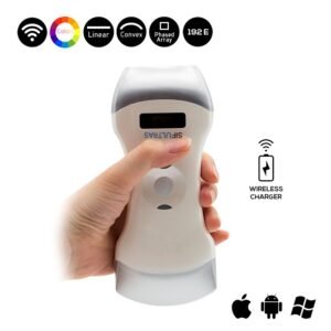
Which ultrasound scanner is best for echocardiography?
Through the movement and rotation of the SIFULTRAS-3.3, you can clearly see the various structures of the heart on the screen. It is an instrument that can dynamically display the structure of the heart cavity, the heart’s pulsation and blood flow.
Cardiac ultrasound is also the only instrument that can visually display valve lesions. Through ultrasound inspection and measurement, doctors can understand whether the valve is functioning properly, whether there is a lesion, the degree of the lesion, and whether to conservative or surgical treatment based on the result of the lesion.
In addition, cardiomyopathy is a disease with an increasing incidence in recent years. Cardiac ultrasound can detect thickening of the myocardium and enlargement of the heart cavity.
For patients with coronary heart disease, ultrasound can visually display the status of myocardial motion (such as the diffuse reduction of left ventricular wall motion) and cardiac function, and indirectly remind clinicians of the possible location of myocardial ischemia.
What diseases are used in the diagnosis and treatment of heart color Doppler ultrasound?
1. Congenital heart disease: For most congenital heart diseases, such as common atrial septal defect and ventricular septal defect, cardiac color Doppler ultrasound has an important role in the diagnosis and does not require other examinations. For some rare and complicated congenital heart diseases, color Doppler ultrasound combined with cardiac magnetic resonance or cardiac catheterization can also confirm the diagnosis;
2. Cardiac valvular disease: such as valve stenosis, insufficiency, prolapse, etc., heart color Doppler ultrasound can clearly see the valve shape, opening and closing status. For rheumatic heart disease, the diagnosis can be confirmed by heart ultrasound only;
3. Cardiomyopathy: such as dilated cardiomyopathy, hypertrophic cardiomyopathy, restrictive cardiomyopathy, etc.;
4. Coronary heart disease: Echocardiography is one of the preferred methods, and the main diagnosis is based on abnormal movement of the myocardial wall in the ischemic area. It also has unique diagnostic value for common complications of coronary heart disease, such as valve insufficiency caused by papillary muscle ischemia, ventricular septal perforation caused by myocardial infarction, ventricular aneurysm, and intracardiac thrombosis. ,
5. Hypertensive heart disease: Mainly observe whether there is hypertension heart damage: such as left ventricular myocardial hypertrophy, left ventricular contraction and diastolic function changes, whether there is secondary coronary heart disease;
6. Pericardial lesions: such as pericardial effusion or constriction;
7. Heart tumor: such as left atrial myxoma;
8. Macrovascular diseases: such as aortic dissection and aortic aneurysm;
9. Others: such as cor pulmonale, infective endocarditis, etc.; in addition to diagnosing the disease, color Doppler ultrasound can also be used for:
Furthermore, a cardiac ultrasound is used for Intraoperative monitoring. During surgery and interventional treatment, transesophageal echocardiography is used to correct and supplement preoperative diagnosis to make up for the shortcomings of transthoracic examination; immediate evaluation of the effect during surgery, such as valvuloplasty and replacement surgery; intraoperative real-time Guide the minimally invasive occlusion process; monitor cardiac function changes during surgery and detect intraoperative complications early. Can improve the quality of surgery, reduce surgical trauma and shorten the operation time.
And of course it is imperative is a postoperative follow-up. To evaluate the surgical effect, recovery of cardiac structure and hemodynamic outcome.


[launchpad_feedback]
Disclaimer: Although the information we provide is used by different doctors and medical staff to perform their procedures and clinical applications, the information contained in this article is for consideration only. SIFSOF is not responsible neither for the misuse of the device nor for the wrong or random generalizability of the device in all clinical applications or procedures mentioned in our articles. Users must have the proper training and skills to perform the procedure with each ultrasound scanner device.
The products mentioned in this article are only for sale to medical staff (doctors, nurses, certified practitioners, etc.) or to private users assisted by or under the supervision of a medical professional.