
Ultrasound-Guided Enlarged Heart Diagnosis
An enlarged heart can be caused by a short-term load on the body, such as pregnancy, or by a medical condition, such as heart muscle weakness, coronary artery disease, heart valve abnormalities, or irregular heart rhythms.
Certain conditions may cause the heart muscle to become thicker or cause one of the chambers of the heart to dilate, making the heart larger. Depending on the condition, an enlarged heart may be temporary or permanent.
Conditions that cause your heart to beat harder than normal or that damage your heart muscle might produce an enlarged heart. For unexplained reasons, the heart might become bigger and weaker. This is referred to as idiopathic cardiomegaly.
A heart condition you’re born with (congenital), to name a few, damage from a heart attack or an abnormal heartbeat (arrhythmia) can cause your heart to enlarge.
In some people, an enlarged heart causes no signs or symptoms. Others may have these signs and symptoms:
· Shortness of breath
· Abnormal heart rhythm (arrhythmia)
· Swelling (edema)
In terms of diagnosis, ultrasound imaging may be used to detect an enlarged heart, which can lead to heart failure, blood clots, or cardiac arrest if left untreated.
For this reason, if you have shortness of breath, an irregular heart rhythm, and swelling in your body, you should undergo an ultrasound.
As may be seen, ultrasound imaging is an important diagnostic technique. The main issue is that not all scanning machines in use throughout the world are functional and professional enough. Doctors and patients should exercise extreme caution when selecting a gadget to ensure clean scan pictures and accurate diagnosis.
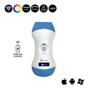
In this context, cardiologists frequently propose the Color Doppler 3 in 1 Wireless Ultrasound Scanner SIFULTRAS-3.31. This wireless ultrasound method has the most promise for detecting Enlarged Hearts. First, it features a cardiac probe, which substantially assists the doctor in evaluating flow direction and velocity in order to properly examine the state of the heart.
Subsequently, this technology allows the doctor/surgeon to immediately see clear scan results of the affected area of the heart, and followingly provides good information on the evolution of the issue and the possible treatment or medicines that should be taken.
What has been said so far should testify that this exclusive device has a superior Color image quality, accurate scan results, is cost-Effective, small and light, easy to carry and operate for evaluation of the critically ill since it exclusively measures volume and percent vascularity when combined with 2D Mode.
All of these high – end features combine to make the SIFULTRAS-3.31 one of the greatest ultrasound scanning machines on which both patients and physicians can trust for an accurate diagnosis, opening the way for quick and efficient treatment of the Enlarged Heart condition.
Reference: Enlarged heart
Disclaimer: Although the information we provide is used by different doctors and medical staff to perform their procedures and clinical applications, the information contained in this article is for consideration only. SIFSOF is not responsible neither for the misuse of the device nor for the wrong or random generalizability of the device in all clinical applications or procedures mentioned in our articles. Users must have the proper training and skills to perform the procedure with each ultrasound scanner device.
The products mentioned in this article are only for sale to medical staff (doctors, nurses, certified practitioners, etc.) or to private users assisted by or under the supervision of a medical professional.