US guided Barbotage for Calcific Tendinitis of the Rotator cuff
Rotator cuff calcific tendinitis is a common condition of unknown aetiology. Hydroxyapatite crystal deposition disease (HADD) is the predominant cause for calcific tendonitis characterized by the deposition of hydroxyapatite (HA) crystals within the intra-articular and periarticular regions of the joint. In around 50% of cases this condition is entirely asymptomatic while 50% of the time being a cause of pain and discomfort. Ultrasound has proven to be very sensitive in detecting these calcific deposits seen as echogenic foci usually accompanied by acoustic shadowing.
Calcific tendinitis effects people between the age of 30 and 40 the most. It is estimated that about 20% of all the shoulder ailments are related to rotator cuff calcification with females being affected more than males. These calcium deposits most commonly involves the supraspinatus tendon and can cause moderate-to-chronic pain and functional disability.
There are various modalities used to treat tendinitis of the rotator cuff including arthroscopic surgery, sonography, shockwave lithotripsy, and ultrasound-guided barbotage (also known as percutaneous aspiration of calcifications).
Barbotage therapy (Often performed in conjunction with a subacromial bursal injection) is an established technique for the treatment of calcific tendinitis. Lavage of the calcifications is usually performed from an anterior approach. It was first described using X-ray machines to guide a needle through the skin to break up the calcification of the rotator cuff. With technological improvements modern ultrasound machines allow a skilled operator to readily ‘see’ the calcification and assess it in 3 dimensions.
This procedure consists of guiding a needle to the location of the calcification through ultrasound guidance. The distal part of the needle is usually not visible within the dense calcium because of posterior acoustic shadowing generated by calcium. Gentle movement of the needle is transmitted to the calcific deposit and observed by ultrasound. Aspiration of calcium is initially attempted. If aspiration is unsuccessful, then needling the lesion can help encourage calcium fragmentation and dispersion. Saline may be injected to help flush out the calcium. Subsequent aspiration may then be performed via the same needle or a second needle may be inserted so that the saline is injected via one needle and aspirated with the other. Extracted calcium is identified in the syringe as solid white gritty material or milk-like fluid. Lavage is usually continued until the aspirate is free of calcific particles but it is not essential to remove the calcium deposit completely.
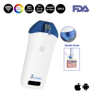
Good-quality ultrasound equipment and a high-frequency (12-15 MHz) linear-array (with a flat surface) probe is required for the efficient treatment of calcific tendinitis and Probe frequency selection depends on the patients build. Based on these parameters we recommend our color doppler mini linear Wi-Fi ultrasound scanner SIFULTRAS-3.51. Using the latest ultrasound probe technology it provides High resolution ultrasound images and High intensity focused digital. It also comes with a needle guide holder to enhance the needle maneuvering Coupled with the software that can quickly locate the depth and diameter of puncture’s navigation.
This procedure is performed by an eligible orthopedist.
Reference: Ultrasound imaging-guided percutaneous treatment of rotator cuff calcific tendinitis: success in short-term outcome
Barbotage is needling of the calcification to trephinate or aspirate the contents of the calcific lesion.
-
Product on saleColor Doppler Mini Linear WiFi Ultrasound Scanner SIFULTRAS-3.51Original price was: $4,500.$2,395Current price is: $2,395.

