US-Guided Cavity analysis And vascular access
In the adult, each kidney is approximately 3 cm thick, 6 cm wide, and 12 cm long. It is roughly bean-shaped with an indentation, called the hilum, on the medial side. The hilum leads to a large cavity, called the renal sinus, within the kidney. The ureter and renal vein leave the kidney, and the renal artery enters the kidney at the hilum. The central region of the kidney contains the renal pelvis, which is located in the renal sinus, and is continuous with the ureter. The renal pelvis is a large cavity that collects the urine as it is produced. Ultrasound is a fundamental device in the diagnosis and treatment of the nephrological patient permitting the analysis of shape, cavities and performance of the kidneys . Nonetheless, the practicality of ultrasound for nephrologists isn’t restricted only to the ultrasound investigation of the kidney but also for cavity analysis, the lower urinary tract and to guide percutaneous techniques, like vascular access for hemodialysis, kidney biopsy and percutaneous nephrostomy or abscess drainage.
Technological development over the past 20 years have lead to high-quality scanners that are portable and affordable. This has greatly expanded the use of point-of-care sonography by clinicians. Nephrologists have been lagging in this area, but an increasing number are incorporating US-guided cavity analysis and vascular access examination into their practice. More importantly training programs are finally starting to meet this need.
nephrology ultrasound investigation requires an ultrasound machines capable of performing studies in two-dimensional and Doppler modes. The equipment should have at least two transducers: a low-frequency convex one for abdominal cavity analysis (kidneys, bladder, prostate, liver-bile duct and abdominal aorta); and a high-frequency linear one for exploration of more superficial structures (pleura-lung, parathyroid glands, carotid and femoral arteries). The ultrasound device should also be able to explore the jugular and femoral veins for cannulation. The same linear transducer will be used in puncture and exploration of the vascular access for HD scanning.
A curved array transducer with a frequencie of 3–6 MHz is used on adult patients, while the pediatric patient should be examined with a linear array transducer with higher frequencies. Artifacts of the lowest ribs always shadow the upper poles of the kidneys. However, the entire kidney can be examined during either normal respiration or breath hold. The kidney will follow the diaphragm and change position accordingly.
Doppler screeninng of the kidney is widely used for vascular access. The vessels are easily portrayed by the color Doppler technique in order to evaluate perfusion. Applying spectral Doppler to the renal artery and selected interlobular arteries, peak systolic velocities, resistive index and acceleration curves can be estimated. For vascular access a meta-analysis by Hind et al compared central venous catheterization using ultrasound-guided puncture with those carried out guided by anatomical markers. They reported a reduction of 86% in relative risk of catheter placement failures using ultrasonography. In addition, a 57% reduction was seen in relative risk of mechanical complications. The mentioned findings are related to punctures of internal jugular veins. these advantages with the adoption of ultrasound guidance proves even more valuable in emergency situation.
Cystic renal masses are portrayed as a distortion of the normal renal structure. Most renal masses are simple cortical cysts with a round appearance and a smooth thin capsule containing anechoic fluid. The incidence increases with age. At least 50% of people above the age of 50 have a simple cyst in one of the kidneys. Cysts cause posterior enhancement as a consequence of reduced attenuation of the ultrasound within the cyst fluid. The simple cyst is a benign lesion that does not require further evaluation. Complex cysts can have membranes dividing the fluid-filled center with internal echoes, calcifications or irregular thickened walls. The complex cyst can be further evaluated with Doppler US. For Bosniak classification and follow-up of complex cysts, either contrast-enhanced ultrasound (CEUS) or contrast-enhanced computed tomography (CT) are used]. The Bosniak classification is divided into four groups going from I, corresponding to a simple cyst, to IV, corresponding to a cyst with solid parts and an 85%–100% risk of malignancy.
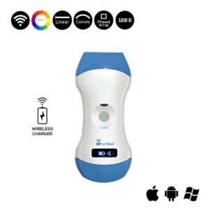
To meet the ultrasound parameters required for these scanning procedurers we recommend the Color Doppler Ultrasound Scanner SIFULTRAS-3.31. This ultrasound scanner features a Convex 3.5/5MHz, Linear 7.5/10MHz making it suitable for both adult and pediatric patients. It’s also equipped with a color doppler for measuring systolic velocity of the renal artery . There is no need to change probe’s head as this can be done by simply changing the software. SIFSOF’s engineer team developed this portable ultrasound machine with nephrology ultrasound diagnosis and intervention in mind. In the application of vascular access, real time ultrasonography achaived by the SIFULTRAS-3.31 means that the progression of the needle to the vessel is done under continuous visualization through this imaging method which has many advantages.
These scanning procedures should be performed by a qualified nephrologist.*
Reference: The role of ultrasound guidance for vascular access
Ultrasonography of the Kidney: A Pictorial Review