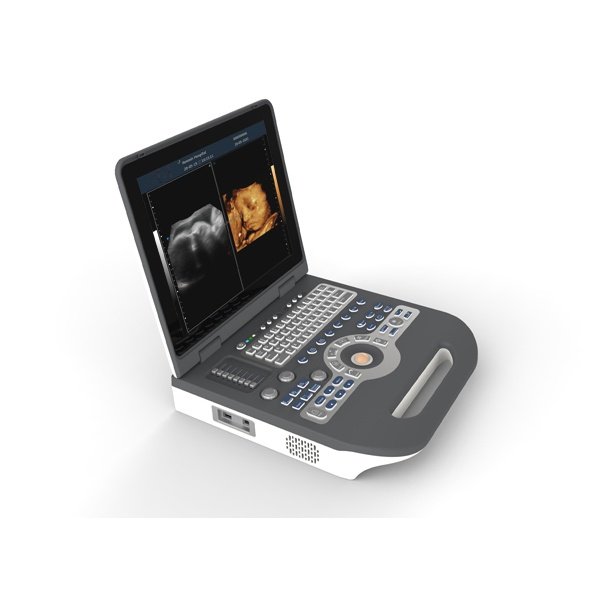


Ultrasound Scanner Notebook Color Doppler SIFULTRAS-8.31
Original price was: $12,000.$8,899Current price is: $8,899.
Convex: R60 (R50) Center frequency 3.5MHz (range: 2-6MHz).
Displayer: 15 inch LCD color display.
Running hours: ≥8h.
Input power: ≤300VA.
Host weight: about 6 kg.
Host appearance size: 370 ×382×90(length × width × height) (mm3).
Two USB port.
S video output port.
For quantity discounts Please call : +1-323 988 5889
10 × Trees planted for one purchased item
![]()
![]()
![]()
![]()
![]()
![]()
![]()
![]()
![]()
![]()
Ultrasound Scanner Notebook Color Doppler SIFULTRAS-8.31
The Notebook Ultrasound Scanner SIFULTRAS-8.31 is based on the high imaging quality of the medical technology products. It is powerful and able to be adapted to many different functions and activitie. This Diagnostic Notebook has a vigorous function with all the optional probes that support his powerness, rich image conversion and adjust package software, clair blood flow image. Not to mention the additional functions like adding PW envelope, mode of three images as well as practical features like real time synchronization. Notebook Ultrasound Scanner SIFULTRAS-8.31with its Classy look and compact is easy to care as it has a portable feature, it has also a hard disk static and dynamic image storage of 120G capacity and 4 Input / output interface: HDMI port, video input/output port, S-VGA, print port, DICOM 3.0,USB port.

With Notebook Ultrasound Scanner SIFULTRAS-8.31 you can measure each organ at a time with detailed specifications. Diagnostic Ultrasound Scanner SIFULTRAS-8.31 has multiple of measurement and analysis : General Measurement, OB and GYN , Cardiac Function, Doppler Blood Flow, Peripheral Blood Vessel, Urology, Orthopedic, Automatic Doppler Flow. Not to mention that the Users can programme protocol numbers, formulas and tables as well as writing Diagnostic report, and add the ultrasound diagnostic image to the report, and print directly.
This Notebook is most used for the examination of the abdomen, Heart, Urology, Obstetrics, Gynecology, Small Organs, Andrology, Musculoskeletal, vascular…

Color Doppler Mode is needed to:
- Give a visual overview of flow within the vessel or heart.
- Rapid identification of vessels, valves, turbulent flow.
- Evaluate flow direction and velocity.
- Measure volume and percent vascularity when combined with 3D Mode.
- Guidance for reproducible quantification of flow velocities using Pulsed-Wave Doppler.
- Locate area of stenosis or thrombosis.
- Determine the existence and amount of arterial plaques and associated turbulent flow.
- Find small vessels such as mouse coronary arteries, femoral and arcuate arteries.
- Evaluate blood flow after a stroke or other cases due to impaired blood flow.
- Observe blood flow to major organs such as heart, kidney, liver pancreas, carotid, abdominal aorta, and others.
This machine is given with a free convex probe and many other optional probes.
The Convex Probe:
Convex probe frequency: 2 – 5 MHZ (multi-frequency, Harmonic frequency ≥5 ), probe scanning angle 20°~85°, visible and adjustable.
The Main Technical Indexes of the Gray-scale imaging mode requirements performance:

The Main Technical Indexes of the color Doppler imaging mode requirements performance:
- Color blood flow image is essential with the gray-scale image of pipes.
- Blood flow direction is identified, no aliasing phenomenon.

The Main Technical Indexes of the Doppler spectrum mode requirements performance:

Other Probes:
- Linear probe frequency: 6.0-12.0MHZ(multi-frequency, harmonic frequency ≥4 ), probe scanningwith trapezoidal imaging technology and 2D beam deflection technology.
- Trans-vaginal probe frequency: 5.0-8.0MHZ(multi-frequency, harmonic frequency ≥2 ), probe scanning angle 20°~160°, visible and adjustable.
- Real time 3D (4D) volume probe frequency: 2.0-5.0MHz, 4 segments multi-frequency.
- Micri-convex probe frequency: 4.0-6.0MHz, 3 segments multi-frequency.
The Main Technical Indexes of the Gray-scale imaging mode requirements performance:

The Main Technical Indexes of the Color Doppler Imaging mode requirements performance:
- Color blood flow image is essential with the gray-scale image of pipes.
- Blood flow direction is identified, no aliasing phenomenon.

The Main Technical Indexes of the Doppler spectrum mode requirements performance:
Pulse wave Doppler mode sampling area cursor position is accurate
Features:
- Full-Digital 2D Gray Scale Imaging.
- Full-Digital Tissue Harmonic Imaging (THI).
- Color Doppler Blood Flow Imaging.
- Directional Color energy Doppler Imaging.
- Pulse Wave Doppler Imaging (PW).
- Space Compound Imaging.
- 2D, Color, Doppler Mode Automatic Optimization Adjustment Technology.
- Real-Time Triple Synchronizing.
- Support 6 Kinds of Language.
- 1 Adaptive Speckle Suppression Technology.
- 1 Hand Free 3D Imaging (Optional).
- 1 Real-Time 4D Imaging (Optional).
- Intelligent Picture – In – Picture Imaging Mode (PIP).
- Monitor: 15 Inch High-Resolution Medical LCD Monitor, Adjustable Angle.
- Probe Connectors: ≥2 Active.
Specifications:
- Convex: R60 (R50) Center frequency 3.5MHz (range: 2-6MHz).
- Displayer: 15 inch LCD color display.
- Running hours: ≥8h.
- Input power: ≤300VA.
- Host weight: about 6 kg.
- Host appearance size: 370 ×382×90(length × width × height) (mm3).
- Two USB port.
- S video output port.
- 2D,B/M,PDI,PW,CFM,4D, imaging mode.
- Gray scale: 256.
- Gray Map: ≥16 level, visible and adjustable.
- Dynamic range: 60-240db(visible and adjustable).
- Resolution: Horizontal≤1mm; Vertical≤0.5mm.
- Under B mode, focus number: 1-6, focus position continuously adjustable.
- STC gain control: ≥8 segments.
- THI: harmonic frequency ≥2 segments.
- Line density: ≥256, visible and adjustable.
- Preset: ≥40 kinds, users can customize the inspection conditions for the optimized images of different organs.
- Max scanning depth: ≥775px, visible and adjustable.
- Scanning angle: 50°-100°, visible and adjustable.
- Cine loop: ≥4800 frames.
- Adaptive speckle suppressio: 0-100 adjustable.
- Amplification: overall amplification, local amplification, M-type amplification(do M-type sampling amplification under both scanning or freeze state).
- Color gain: adjustable.
- Color frequency: ≥3 kinds, visible and adjustable.
- Sampling frame: size and position adjustab.
- PW blood flow measurement speed: min speed: ≤0.2 cm/s, max speed: ≥37500px/s.
- PW Doppler frequency: ≥3 kinds, CW Doppler frequency: ≥15 kinds, visible and adjustable.
- Real-time automatic Doppler envelope mapping and automatic measurement and analysis.
Applications:
This Notebook is most used for the examination of the abdomen, Heart, Urology, Obstetrics, Gynecology, Small Organs, Andrology, Musculoskeletal, vascular…
Doctor Specialities:
SIFULTRAS-8.13 is used by MSK doctors, Cardiyologists, Gastroenterologists, Gynecologists, GO Doctors, Neonatologists, Nephrologists, Obstetricians, Pediatricians, Radiologists, Urologists, Surgeons, Vascular Doctors, Andrologists…
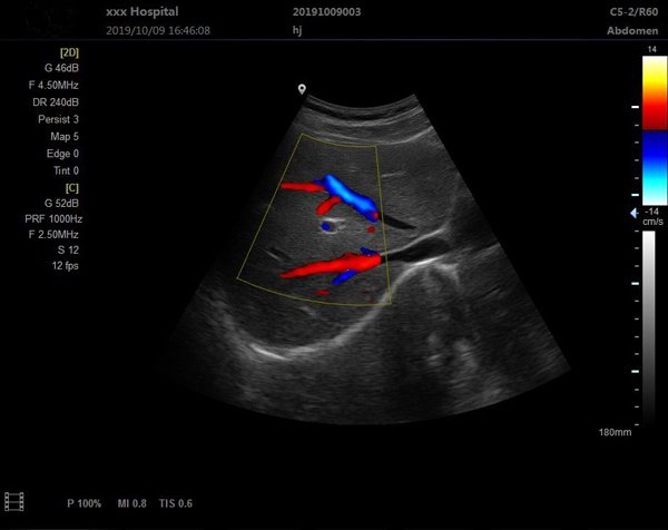
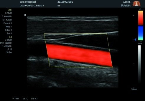
Scan Results
Certifications:

Color Doppler Convex Diagnostic Ultrasound Scanner SIFULTRAS-8.31 (convex probe)
Related products
-
Aesthetic Plastic Surgery
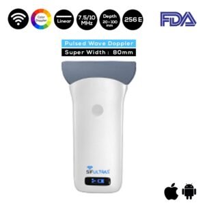
PW Doppler Wireless Ultrasound Scanner 7.5-10 MHz Super Width Linear Probe SIFULTRAS-5.39
Original price was: $5,000.$2,895Current price is: $2,895. Add to cart -
Health Connected Devices
Color Ophthalmic A-Scan Ultrasound Scanner SIFULTRAS-8.21
Original price was: $3,000.$2,350Current price is: $2,350. Add to cart -
Cardiologist
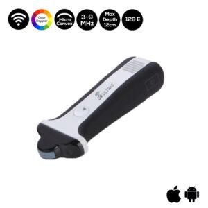
Micro Convex Ultrasound Scanner Color Doppler Wireless SIFULTRAS-5.03
Original price was: $4,000.$3,199Current price is: $3,199. Add to cart -
Anesthesiologist
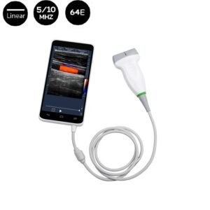
Color 5-10 MHz WiFi/Bluetooth Linear 64E Ultrasound Scanner SIFULTRAS-3.0
Original price was: $2,970.$2,145Current price is: $2,145. Add to cart