
Neuroma Injection Ultrasound Scanner
Different types of nerve damage can result in neuromas anywhere in the body. They are a component of the reparative process, and the presence of a distal tract or Schwann cell determines their appearance
. The location of the neuroma may occasionally be painful. With residual limb pain, the source of the pain may be due to ischemia, which is caused by a drop in blood flow, scar tissue producing traction on the nerve and neuroma, or it may be caused by compression by the surrounding tissues or vessels. It is difficult to control the neuroma’s pain. Local anesthetic, steroid, and neurolytic drug injections, cryo-ablation, radiofrequency ablations, and surgical revision are among the therapy possibilities Unfortunately, changing the surgical procedure to stop neuromas from forming hasn’t always worked.
Based on how they appear, neuromas fall into two main groups. Spindle neuromas develop due to microtrauma brought on by friction or irritation to the nerve, which is not severe enough to disrupt the trunk. Following severe damage that results in the nerves being surgically or traumatically sectioned, terminal neuromas can develop. The initial diameter of the nerve, the number of damaged axons, recurring trauma or irritation, the environment, and the rate of growth all affect the size of the neuromas. A neuroma typically develops over the course of 6 to 8 weeks. After a year, neuromas typically stop growing.
What is the most suitable ultrasound for Neuroma Injection?
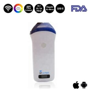
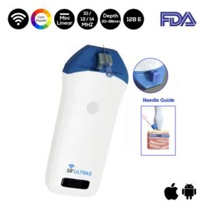
A scout scan is carried out using a linear array transducer with the proper frequency, often in the range of 10–14 MHz, such as the Color Doppler Mini Linear WiFi Ultrasound Scanner SIFULTRAS-3.51 , depending on the depth of the target, depending on the location of the pain and neuroma. The scan is carried out in both transverse and longitudinal views to determine the neuroma’s potential location. The nerve is tracked proximally and distally after the neuroma has been detected in order to determine its length and breadth. Without a color flow Doppler analysis to pinpoint the nearby vessels, no scan is complete. This makes it easier to plan a needle trajectory.
Reference: Ultrasound-Guided Neuroma Injection – ASRA
Disclaimer: Although the information we provide is used by different doctors and medical staff to perform their procedures and clinical applications, the information contained in this article is for consideration only. SIFSOF is not responsible neither for the misuse of the device nor for the wrong or random generalizability of the device in all clinical applications or procedures mentioned in our articles. Users must have the proper training and skills to perform the procedure with each ultrasound scanner device.
The products mentioned in this article are only for sale to medical staff (doctors, nurses, certified practitioners, etc.) or to private users assisted by or under the supervision of a medical professional.