
Ultrasound-Assisted Joint Effusion
An increase in fluid volume within the synovial compartment of a joint is referred to as an effusion. Only a little amount of physiological intra-articular fluid is often present. Exudate, transudate, blood, and/or fat may accumulate abnormally as a result of trauma, inflammation, infection (such as pus), or exudate and transudate. Intentional injection of contrast material into the joint space during an arthrogram causes an iatrogenic effusion.
An instance of lipohemarthrosis is when there is an intra-articular fracture and there is a fat-fluid level because marrow fat leaks into the joint through the fracture. Fat will float to the surface and appear as a “fat-fluid” level on top of the blood on any radiography with the horizontal beam parallel to the level because it is less dense than blood. Although lipohemarthroses can develop in other joints (such as the shoulder), they are the easiest to spot in the knee.
It might be challenging to spot joint effusion on conventional radiographs, especially for non-radiologists. Understanding the usual manifestations and symptoms of joint effusions might help in diagnosis.
Which ultrasound Scanner is suitable for joint effusion?
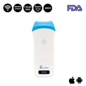
The Linear Wireless Ultrasound Scanner SIFULTRAS-5.34 – Color Doppler is highly recommended for this procedure. The quadriceps tendon can be seen in the near field and the cortex of the femur can be seen in the far field after placing the probe just cephalad to the patella. The suprapatellar recess of the knee joint, which intercalates between the suprapatellar and prefemoral fat pads, is located between these two structures. Visualization of the lateral and medial joint recesses is possible with caudad angulation. A fluid effusion will enlarge the visible crevices; this fluid may be anechoic or have mixed echogenicity. Internal color flow is absent Doppler signals should be discernible, and probe compression should cause a change in the collection’s structure or internal movement
Reference: Joint effusion


Disclaimer: Although the information we provide is used by different doctors and medical staff to perform their procedures and clinical applications, the information contained in this article is for consideration only. SIFSOF is not responsible neither for the misuse of the device nor for the wrong or random generalizability of the device in all clinical applications or procedures mentioned in our articles. Users must have the proper training and skills to perform the procedure with each ultrasound scanner device.
The products mentioned in this article are only for sale to medical staff (doctors, nurses, certified practitioners, etc.) or to private users assisted by or under the supervision of a medical professional.