
The Superficial Fascia of The Abdomen
The layers of the abdominal wall consist of the skin, superficial fascia, and muscles. The superficial fascia of the abdomen which forms the greatest part of the abdominal wall consists of a single layer containing a variable amount of fat; but near the groin it is easily separated into two layers, between which are found the superficial vessels and nerves and the superficial inguinal lymph glands.
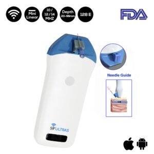
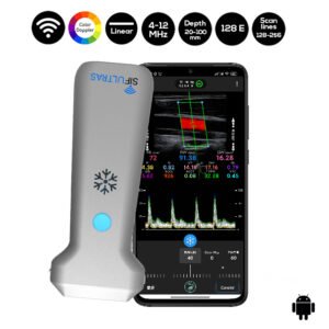
Which ultrasound scanner is used for Superficial Fascia of the Abdomen imaging?
Ultrasound with high resolution and frequency SIFULTRAS-3.5 is strongly suggested in these medical operations. As a high frequency probe offer superficial images, which is where The Superficial Fascia of The Abdomen is.
Using ultrasonography, fibrous connective tissue layers appear as echogenic and echolucent bands. Ultrasonography has the biggest advantage of assessing the sliding between the various fascial layers in real time and permitting the accurate measurement of their thickness.
Ultrasound imaging is being greatly used by Physical and Rehabilitation Medicine (PRM) specialists to measure the thickness of abdominal muscle.
For healthy person, doctors need to position The probe along the right lateral abdominal wall at the height of the 12th rib : above the umbilicus at the linea alba, to the side of and approximately 2 cm from the umbilicus, along the mammillary line and along the anterior axillary line.
Each rater should measure about 17 anatomical structures six times during two sessions. Doctors admit that the reliability of using ultrasound in this procedure is good under both conditions for the fasciae and excellent for the muscles.
Knowledge of the fascial anatomy of the abdominal wall is required for detailed ultrasound examinations and for ameliorating reliability.
These results prove that ultrasound imaging is a reliable, non-invasive, cost-effective instrument for assessing the abdominal muscles/fasciae.
This procedure is also performed by for orthopedic specialists, experts on fasciae, PRM resident, PRM specialists..

[launchpad_feedback]
Although the information we provide is used but doctors, radiologists, medical staff to perform their procedures, clinical applications, the Information contained in this article is for consideration only. We can’t be responsible for misuse of the device nor for the device suitability with each clinical application or procedure mentioned in this article.
Doctors, radiologists or medical staff must have the proper training and skills to perform the procedure with each ultrasound scanner device.