
Hysterosonography
Hysterosonography or infusion Sonohysterography is the evaluation of the endometrial cavity using the transcervical injection of sterile fluid. The primary goal of Hysterosonography is to visualize the endometrial cavity in more detail than is possible with routine endovaginal sonography.
This technique may also be used to assess tubal patency. An increase in the amount of free pelvic fluid at the end of the procedure indicates that at least one tube is patent.
Which ultrasound scanner is best suited for Hysterosonography?
Hysterosonography is usually conducted with a high-frequency endovaginal transducer SIFULTRAS-5.43. In cases of an enlarged uterus, additional transabdominal images during infusion may be required to fully evaluate the endometrium. The transducer should be adjusted to operate at the highest clinically appropriate frequency under the ALARA (as low as reasonably achievable) principle.
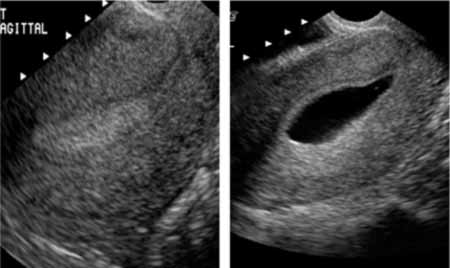
The procedure involves injecting sterile saline into the uterus, distending or enlarging the uterus. The saline outlines the lesion and allows for easy visualization and measurement.
Precatheterization images should be obtained and recorded, in at least 2 planes, to show normal and abnormal findings. These images should include the thickest bilayer endometrial measurement, which includes the anterior and posterior endometrial thicknesses, obtained in a sagittal view.
Once the uterine cavity is filled with fluid, a complete survey of the uterine cavity should be performed and representative images obtained to document normal and abnormal findings. If a balloon catheter filled with saline is used for the examination, images should be obtained at the end of the procedure with the balloon deflated to fully evaluate the endometrial cavity, particularly the cervical canal and lower portion of the endometrial cavity.
After cleansing the external os, the cervical canal and/or uterine cavity should be catheterized using aseptic technique, and appropriate sterile fluid should be instilled slowly by means of manual injection under real-time sonographic imaging. Imaging should include real-time scanning of the endometrial and cervical canal.Imaging may include evaluation of fallopian tube patency if indicated.
Color Doppler sonography may be helpful in evaluating the vascularity of an intrauterine abnormality and tubal patency. Three-dimensional imaging, in particular reconstructed coronal plane imaging, is useful in the assessment of Mullerian duct anomalies and for preoperative mapping of myomas.
This minimally invasive ultrasound technique creates pictures of the inside of a woman’s uterus.
This technique can also be valuable for evaluating unexplained vaginal bleeding that may be the result of uterine abnormalities such as:
- Congenital defects.
- Masses.
- Adhesions (or scarring).
- Polyps.
- Fibroids.
- Atrophy.
This test can be done in your obstetrician–gynecologist’s (ob-gyn) office, a hospital, or a clinic. It usually takes less than 30 minutes.
Refreence : AIUM Practice Parameter for the Performance of Sonohysterography.


[launchpad_feedback]
Although the information we provide is used but doctors, radiologists, medical staff to perform their procedures, clinical applications, the Information contained in this article is for consideration only. We can’t be responsible for misuse of the device nor for the device suitability with each clinical application or procedure mentioned in this article.
Doctors, radiologists or medical staff must have the proper training and skills to perform the procedure with each ultrasound scanner device.